MicroED Core
MicroED Core Services
The microcrystal electron diffraction core at UCLA
The MicroED Core
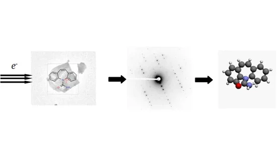
The MicroED Core is a cryo-EM facility based at UCLA’s David Geffen School of Medicine, dedicated to make MicroED structure solution accessible to academia and industry. Services include all steps from sample mounting to refined structure, according to your needs and in order to suite your workflow.
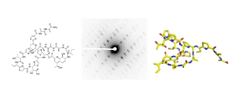
The Core specializes in small, medium, or macromolecular structure determination using MicroED. The technique has a growing success record with a large variety of compounds, including chemical substances, peptides and biosynthesized natural products, membrane-bound proteins, and large biological complexes.
What MicroED will do
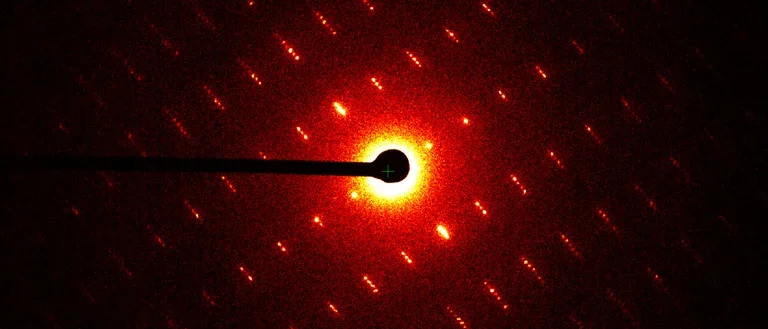
MicroED is suitable for any well-diffracting crystal. It has established itself as a method in its own right, but can also fill a complementary role to X-ray crystallography, NMR techniques, or single particle analysis. As crystals need only be between tens to a few hundreds of nanometers in size, MicroED has proven essential in cases where larger crystals are hard to obtain for a variety of reasons, for instance because re-crystallization is difficult or time consuming, or only small amounts of sample are available. Examples include membrane proteins known to produce smaller crystal and assemblies of larger molecules that also frequently result in limited crystal sizes. Despite the need for only nano-sized crystals, the resolutions achieved are often comparable to those obtained by X-ray crystallography.

About us
The MicroED Core was launched in 2020. Its staff employs state-of-the-art protocols on cutting-edge hardware to manage your project through the structure solution process. We are actively following developments in MicroED and new software and technologies are constantly added.
The Core uses a Talos Arctica microscope running at 200 keV with a high-precision rotating stage. The Core has all necessary laboratory accessories needed to perform MicroED.

Services
For Academia and Industry
Services include all steps toward a refined structure as needed, including sample mounting, initial screening and diffraction analysis, data collection, structure solution, and model refinement. Any additional work can be performed as requested on an hourly payment basis. Although the technique shares fundamental concepts with X-ray crystallography, there are differences, and the staff will assist and advise with their expert knowledge throughout the entire process to ensure the best outcome.
As a crystallographic technique, the basic requirement is that the sample exhibits some crystalline properties. Crystals in water solution are applied directly to the grid and frozen at cryogenic temperatures for data collection. Powder or grainy samples are usually applied to an electron microscopy grid and scanned in the electron beam, which will show the diffractive properties of each individual crystal. The sample need not be homogenous nor are the crystallographic properties required to be identical across the sample. If diffracting crystals are found, data can be collected.
All customers, please contact us for a quote.
MicroED Core Staff


